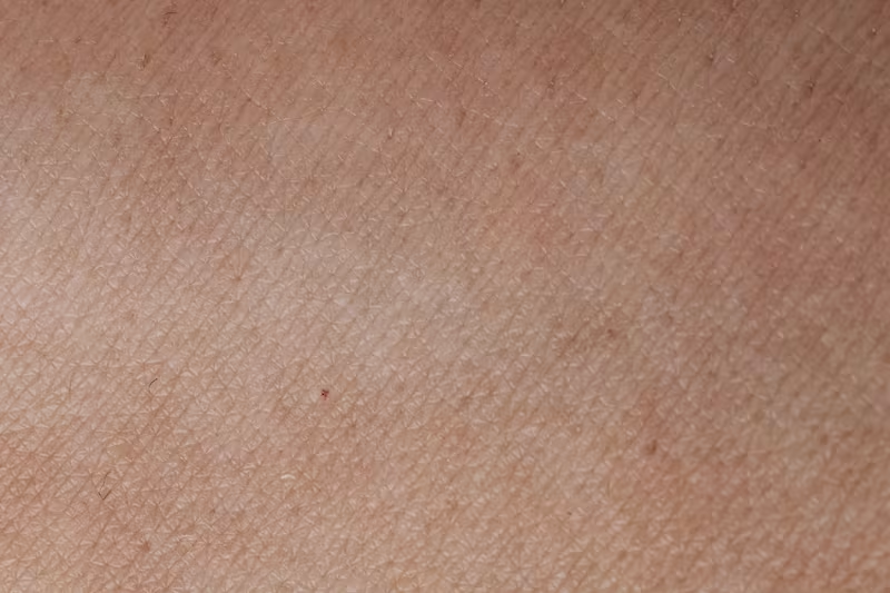
Dermatology lends itself naturally to machine learning because so much diagnostic reasoning is visual. Atypical moles, evolving lesions, subtle color changes, scaling patterns, and border irregularities all carry clinical meaning. These patterns can be learned by an AI system, but only if the training images are labeled with precision.
In dermatology AI, medical image segmentation is not a technical afterthought, it is the foundation. Weak annotation produces brittle models. Strong annotation, performed with clinical context, produces reliable diagnostics, equitable performance across skin tones, and tools that dermatologists can trust in real practice.
Organizations such as the American Academy of Dermatology (AAD) and DermNet have long emphasized how visual interpretation relies on nuance. AI systems must learn those nuances too. That learning starts with annotated data.
What Image Annotation Means in Dermatology
At its core, image annotation is the process of labeling structures or skin patterns within images so AI models can learn what each region represents. In dermatology, annotation often captures more than a single object, it encodes clinical significance.
Annotations may include:
- Lesion contours, capturing irregular borders, asymmetry, or pigment variation
- Localization tags, such as “scalp,” “back,” or “acral surface,” which influence differential diagnoses
- Disease classification, including benign nevi, melanoma, psoriasis, actinic keratosis, eczema, or basal cell carcinoma
- Severity or stage indicators, especially important in chronic inflammatory diseases
- Metadata such as skin tone, using standards like the Fitzpatrick scale
- Pixel-level segmentation, especially when distinguishing subtle lesion features
These annotations help train models for classification, segmentation, anomaly detection, and progression monitoring across diverse dermatology use cases.
Why Dermatology AI Depends on Expertly Annotated Images
AI models can only recognize patterns they have been explicitly taught. Without expert annotation, a model may misinterpret visual noise as pathology or overlook subtle but clinically important features.
Training reliable diagnostic systems
Annotated dermatology images allow models to learn:
- How to differentiate melanoma from benign lesions
- How color, texture, and border irregularity influence diagnosis
- When rashes reflect infection, inflammation, allergy, or systemic disease
- How to separate skin from clothing, hair, or background distractions
High-quality annotation is especially important for dermoscopic images, where microscopic patterns, dots, streaks, pigment networks, carry diagnostic value. Many landmark dermatology AI studies published via NCBI demonstrate that model accuracy improves dramatically when annotations are dermatologist-reviewed.
Reducing bias across skin tones
Bias in dermatology AI has been widely documented. Studies show that models trained overwhelmingly on lighter skin underperform for darker skin tones, increasing diagnostic disparities.
To counter this, annotations must reflect full variations in Fitzpatrick I–VI skin types. Datasets like Fitzpatrick17k and the ISIC Archive demonstrate how diverse representation leads to measurable improvements in model performance.
Enabling clinically meaningful validation
Annotations form the ground truth for evaluating model accuracy. In dermatology, this often requires dermatologist review or biopsy-confirmed cases. Annotations form the ground truth for evaluating performance. AUC-ROC, sensitivity, and Dice metrics used in dermatology AI research—such as those reported by the National Institutes of Health depend on accurate labeling.
How Dermatology Annotation Powers Real Clinical and Commercial Applications
Dermatology is among the fastest-evolving AI fields. Precise annotation unlocks a wide spectrum of applications in both clinical practice and digital health.
Skin cancer detection and melanoma triage
Melanoma remains one of the most dangerous skin cancers, but early detection dramatically improves survival rates. AI systems trained on annotated clinical and dermoscopic images can:
- Flag suspicious lesions
- Prioritize high-risk cases for urgent review
- Support triage in clinics and primary care
- Power mobile screening tools for early detection
Apps like SkinVision and research tools highlighted by the World Health Organization emphasize how annotation quality directly influences algorithmic sensitivity and specificity.
Teledermatology and remote clinical assessment
As telemedicine expands, dermatology is becoming increasingly digital. Annotated images are used to train models that support:
- Automated triage for remote consultations
- Prioritization of severe conditions
- Recognition of common diseases like eczema, acne, fungal infections, and urticaria
- Cross-skin-tone performance in multicultural regions
Platforms such as First Derm and Tesserae Health rely on well-annotated datasets to ensure safe diagnostic guidance for diverse patient populations.
Dermatopathology and histology interpretation
Whole-slide images (WSI) offer another layer of dermatological insight. Annotators label:
- Tumor boundaries
- Epidermal and dermal structures
- Inflammatory patterns
- Atypical cell clusters
Dermatopathology AI, including research projects cited by the NIH, requires pixel-precise masks and expert consensus to reach clinical reliability.
Monitoring chronic skin diseases
Conditions like psoriasis, vitiligo, rosacea, and atopic dermatitis evolve over time. Annotated time-series images enable AI models to:
- Estimate severity scores
- Track lesion expansion or regression
- Quantify pigment changes
- Evaluate treatment response
This is particularly valuable for clinical trials, telemedicine follow-up, and personalized dermatology.
Aesthetic and cosmetic dermatology
Beyond clinical care, AI helps clinics and skincare brands analyze:
- Wrinkles and fine lines
- Pore visibility
- Acne severity
- Pigmentation patterns
- Facial symmetry
These models rely on granular annotation to produce consistent recommendations and simulations for patients.
Building High-Quality Dermatology Datasets
The performance of dermatology AI systems is directly tied to how well their datasets are curated and annotated. Strong datasets share several characteristics.
Diversity across skin tones and demographics
Bias often emerges when datasets disproportionately represent lighter skin. A balanced dataset includes:
- All Fitzpatrick skin types
- A broad range of ages
- Varied anatomical locations
- Images captured under different lighting and devices
Training with this diversity measurably improves fairness and real-world accuracy.
Clinically meaningful labels, not generic tags
Dermatology is nuanced. Instead of simply labeling a lesion “psoriasis,” annotations may include:
- Subtype (plaque, inverse, pustular, guttate)
- Severity (mild, moderate, severe)
- Anatomical location
- Coexisting findings (scaling, erythema, excoriation)
These structured annotations enable more personalized AI outputs.
Multi-stage annotation and clinical review
A robust dermatology workflow often includes:
- Initial labeling by trained technicians
- Medical review by dermatologists
- Consensus resolution for complex cases
- Final QA and dataset versioning
This ensures that clinical subtleties are accurately captured, especially in conditions where visual ambiguity is common.
Privacy, consent, and ethical sourcing
Dermatology images contain sensitive health information. Datasets must comply with GDPR, HIPAA, and local regulations, with attention to:
- Informed patient consent
- Removal of identifiable features
- EXIF metadata cleaning
- Secure storage and access control
Ethical compliance is foundational, especially when datasets include patient-captured mobile images.
Challenges Unique to Dermatology Image Annotation
Dermatology introduces complexities not always present in other clinical imaging fields.
Visual variability across conditions
Lesions may appear differently depending on skin type, lighting, age, or disease stage. Consistent labeling requires annotators to recognize these variations.
Limited availability of expert annotators
Dermatology expertise is specialized. Annotators without medical training may mislabel similar-appearing conditions. Dermatologist review is often essential.
Subjectivity in classification
Even experts may disagree visually. Some lesions require biopsy for confirmation. Annotation pipelines must incorporate:
- Consensus systems
- Clinician feedback loops
- Clear visual labeling guidelines
Regulatory and privacy constraints
Dermatology images often include more identifiable features than radiology or pathology, requiring stricter privacy management.
How AI Models Learn from Dermatology Annotations
Once the dataset is annotated, models can be trained using architectures such as CNNs, Vision Transformers, and hybrid multimodal networks.
Dermatology models often learn:
- Lesion classification and triage
- Lesion localization
- Border delineation
- Disease progression modeling
- Uncertainty estimation to flag ambiguous cases
The better the annotations, the more reliably the model generalizes to real clinical environments.
Emerging Trends That Will Shape Dermatology AI
Dermatology is one of the most active frontiers for medical AI research. Several trends are accelerating progress.
Foundation models trained on dermatology images
Large vision-language models like CLIP and MedCLIP are being adapted for dermatology. These models can understand images alongside patient descriptions (“itchy,” “growing,” “painful”), improving triage accuracy and accessibility.
Federated learning across hospitals
Instead of sharing patient data, hospitals share encrypted model updates. This protects privacy while improving performance across diverse populations.
Synthetic skin images
GANs and diffusion models can generate realistic dermatology images, particularly for rare diseases or underrepresented skin tones. These synthetic data examples help balance datasets but must be clearly flagged.
Explainable dermatology AI
Tools such as Grad-CAM and SHAP help dermatologists understand why a model made a specific prediction, an essential step toward clinical trust and regulatory approval.
On-device dermatology models
Mobile processors increasingly support real-time analysis directly on smartphones. This unlocks offline triage for regions with limited connectivity and enhances patient privacy.
Evaluating Dermatology AI Models
Performance is measured using metrics such as:
- Sensitivity and specificity
- Precision and recall
- Dice coefficient for segmentation
- AUC-ROC for binary classification
- Clinical validation studies comparing AI to dermatologist performance
Technical and clinical metrics must both be met for safe deployment.
Looking Ahead: AI as a Partner, Not a Replacement
Despite rapid progress, AI will not replace dermatologists. Instead, it will:
- Accelerate triage
- Improve screening reach
- Enhance diagnostic confidence
- Reduce clinician workload
- Support global health efforts
Dermatologists bring clinical judgment, patient counseling, and contextual reasoning, capabilities that AI complements but cannot replace.
Partner with DataVLab for Dermatology AI
High-quality annotation determines whether a dermatology AI model performs reliably or fails in real conditions. At DataVLab, we specialize in clinically informed, high-precision annotation workflows tailored to dermatology:
- Lesion segmentation
- Dermoscopic pattern labeling
- Multi-stage expert review
- Diverse skin tone datasets
- Secure, compliant processes
Your dermatology AI deserves expert-level data quality.
If you’re building diagnostic tools, teledermatology platforms, or research datasets, we can support you from the first batch of images to full-scale production.
👉 Ready to strengthen your dermatology dataset? Contact DataVLab today to discuss your project.


